The objectives of this clinical case are to discuss the normal anatomy of the patellofemoral joint and the different therapeutic options in case of an inveterate patellar dislocation.
- A 31-year-old male patient
- Knee trauma when he was 10 years old
- Failed soft tissue surgery 20 years ago
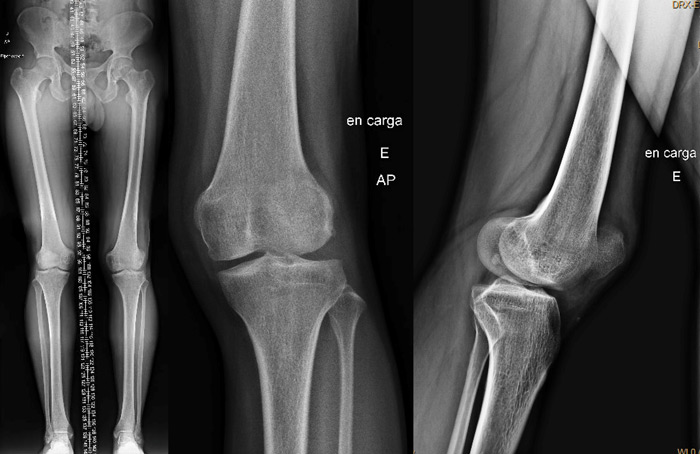
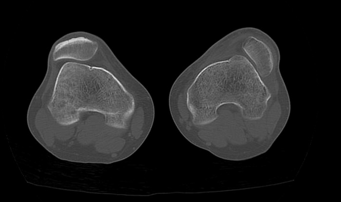
- On physical examination, he presented with a non-reducible patellar dislocation which causes pain and functional loss
Pre-operative X-rays
Pre-operative CT-scan
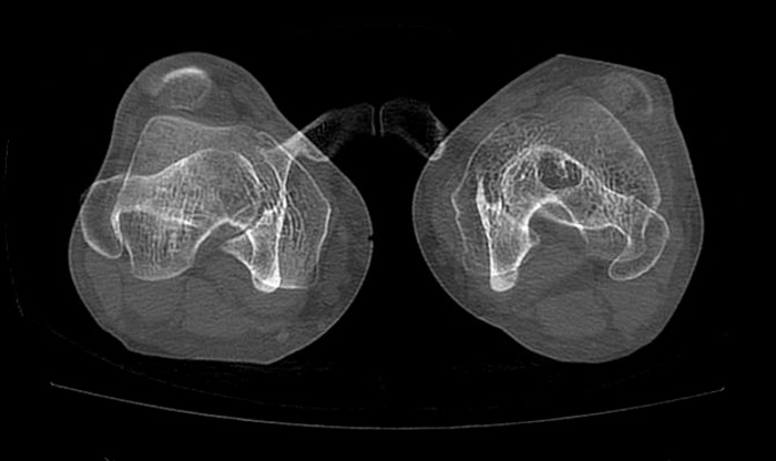
Pre-operative measurements
- HKA = 180°
- TT-TG = 24 mm
- Femoral neck anteversion (FNA) = 27.5°
- Dejour type C trochlear dysplasia
What are the normal values of the femoral neck anteversion and the TT-TG?
- ✔️ FNA = 15, TT-TG = 9
Scorcelletti M, Reeves ND, Rittweger J, Ireland A. Femoral anteversion: significance and measurement. J Anat. 2020 Nov
Rhee, SJ., Pavlou, G., Oakley, J. et al. Modern management of patellar instability. International Orthopaedics (SICOT) 36, 2447–2456 (2012)
How would you manage this condition?
- ✔️Tibial tubercle medialisation
- ✔️Femoral derotation osteotomy
- ✔️Lateral patellar release
- ✔️MPFL reconstruction
Final strategy decision
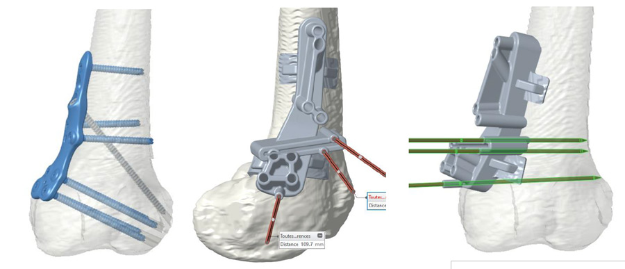
The patient presented with an increased femoral anteversion of 14° in comparison to his contralateral lower limb, and an increased TT-TG of over 20°.
We usually leave the trochleoplasty as the last surgical option. Our choice was then to perform...
1 - a femoral derotation osteotomy of 14° thanks to a personalized cutting guide (PSI-NEWCLIP).
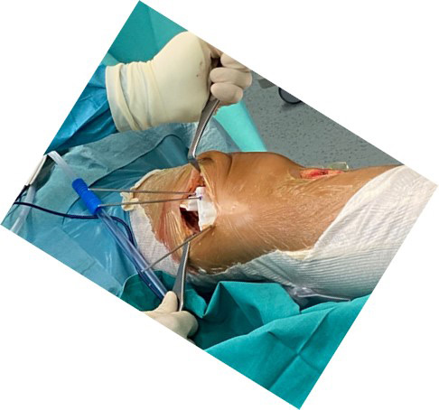
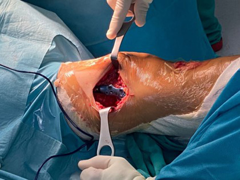
2 - a tibial tubercle medialisation according to the Elmslie – Trillat technique, a TT-TG over 20 should be medialised
Cox JS. An evaluation of the Elmslie-Trillat procedure for management of patellar dislocations and subluxations: a preliminary report. Am J Sports Med. 1976
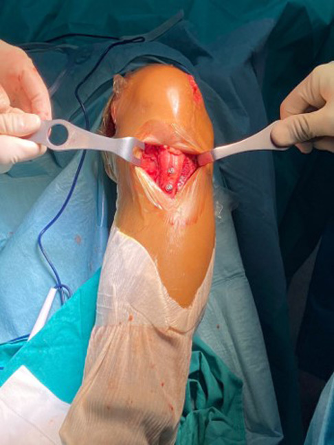
3 - a lateral release
4 - an MPFL reconstruction using a gracilis tendon.
Ahmad R, Jayasekera N, Schranz P, Mandalia V. Medial patellofemoral ligament reconstruction: a technique with a "v"-shaped patellar tunnel. Arthrosc Tech. 2014 Sep 22
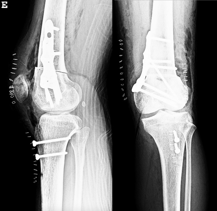
Post-operative X-rays


Comments
(No subject)
In order to perform a better surgical plan I need perfect lateral view x-rays (CT Scan show more D than C trochlear Dysplasia; and no Caton-Deschamp (C/D) index mesurements).Usually flexion patella dislocation is associated with short extensor mechanism (so the choose between TTO and FDO +\- lenthening of extensor mechanism is related to C/D)